
The Four O'Clock Window
At four in the afternoon the staining line is quiet and the bench is not. Racks of finished slides sit in open air, waiting for someone to coverslip them. The stain is done — hematoxylin sharp, eosin clean — and now the slides wait, five minutes or fifty, while the staff clear the backlog at embedding. Nothing looks wrong. The slide still shows what it showed at the end of the line. But every minute of open-air waiting taxes the work that just finished: dust settles, solvent breathes off, and the section begins to dry in patches. Coverslipping is the frame around the picture. In most labs the frame is added later, by hand, in batches — and the gap in between is the part nobody owns.
The Window Nobody Owns
Take a slide through its last steps: deparaffinize, stain, dehydrate, clear, coverslip. After the clearing bath the slide must be protected while it is still wet with solvent — that is what the mounting step is for. Between that bath and the coverslip, the section is at the mercy of the room.
In my experience the delay is rarely one long wait. It is a series of small ones. A rack finishes and sits until someone carries it. The carries happen in bursts, between other jobs. Each slide in the rack waits a different amount of time, which means the same batch is processed differently: the first slide is covered in minutes, the last an hour later. Dust and fibres settle on wet sections. Solvent evaporates from the tissue surface, and a section that dries before mounting picks up unevenness that no one notices until it is under the scope. The work that took an hour to stain is judged in the ten minutes after it was left alone.
Most of what lands on a waiting slide is not dramatic. A fibre from a lab coat, a film of dust, a thumbprint at the edge of the section. Under the scope these read as noise the pathologist has to look past, and when drying has gone further — a patch that never rehydrated evenly — the slide comes back for re-staining. The re-stain is rarely counted as a defect in the stain; it is counted as staff time, reagent time, and a report that leaves the lab a day late. In my experience, the lab that tracks its re-stains can usually trace a good share of them to this uncovered stretch rather than to the staining program itself.
Two Architectures
There are two ways to build this part of the lab. The common one is separate machines and a pair of hands between them: the stainer finishes a rack, a technician lifts it out, carries it across the room, and loads the coverslipper. Every carry is a timing decision made by a person who is also answering the phone.
The other way is an integrated workstation — the staining line and the coverslipper built as one machine, with the finished slide handed from one to the other automatically. No rack leaves the process, no open-air wait, no decision about when the next step happens. The slide that finishes staining is the slide that gets covered, in order, first in first out.
This is why an integrated line uses film rather than glass coverslips. A staining line that runs hundreds of slides an hour needs a coverslipper that can match its rhythm — and a film coverslipper finishes in seconds, with nothing to cure overnight and no adhesive overflow to wipe. Work published in the Journal of the American Society of Cytopathology compared glass, film and liquid coverslipping and found film the fastest route from staining to a scannable slide. Glass coverslips vs film: what I specify instead explains the rest of that decision. Read the coverslipping methods study
Matching the Beat
If you design this line, the coverslipper has to outrun the stainer, not keep pace with it. Staining arrives in bursts — a rack ends, a protocol finishes, a frozen case lands in the middle of a routine run. The coverslipper is the last step of the day, and the one place a backlog can form that keeps staff in the lab after hours. Give the last step headroom and the backlog disappears before it starts.
The same logic applies to protocols. A histology lab does not run one stain. It runs H&E in the morning, Pap smears for cytology, frozen sections whenever the phone rings. A workstation that runs several protocols at once, in parallel, keeps those streams separate instead of forcing the lab to finish one before starting another. When the line can finish a day's routine work and then run unattended, the evening shift stops being a staffing problem.
Think about the end of the day. The last rack of a run is also the rack nobody wants to wait for: the technician is watching the clock, and the coverslipper is the last machine between the lab and the parking lot. A line whose final step runs faster than its middle steps clears that rack before a backlog forms — and a line that can hold a full run's finished output lets the machine work past five o'clock on its own.
First Slide to Last Slide
The quietest quality problem in staining is drift. The first rack of the morning looks right; the fourth rack is paler, or the counterstain has shifted, and slides start coming back to be re-stained. In my experience most re-stains trace to two causes: reagent age and timing. Hematoxylin oxidizes as it works. Alcohols pull water from the air. A program that was correct in January is not the same program in June unless someone is watching the reagents, not just the clock.
The pattern is predictable if you watch for it. A protocol that produces crisp nuclei on the first rack of fresh reagents produces softer ones as the hematoxylin works through the day, because dye uptake falls as the stain exhausts. Eosin drifts the other way as the alcohol in its bath dilutes. Same program, same clock, different chemistry — which is why a lab that re-stains rarely re-stains the first rack. In my experience the re-stained slides cluster in the afternoon, when the day's reagent load has caught up with the morning's settings.
Removing the human hand removes the timing drift — automated staining is measurably more uniform within a batch than manual staining, as an IEEE conference study of automated stainers showed. What an automatic H&E stainer cannot remove is reagent aging, because that is chemistry. The answer is a machine that watches the reagents themselves and adjusts the protocol while the run is happening — which is what the product section below is about. Why Monday-morning H&E looks wrong (usually not the machine) covers the diagnostic side of the same story. Automated vs manual staining uniformity (IEEE study)
What We Built to Answer These
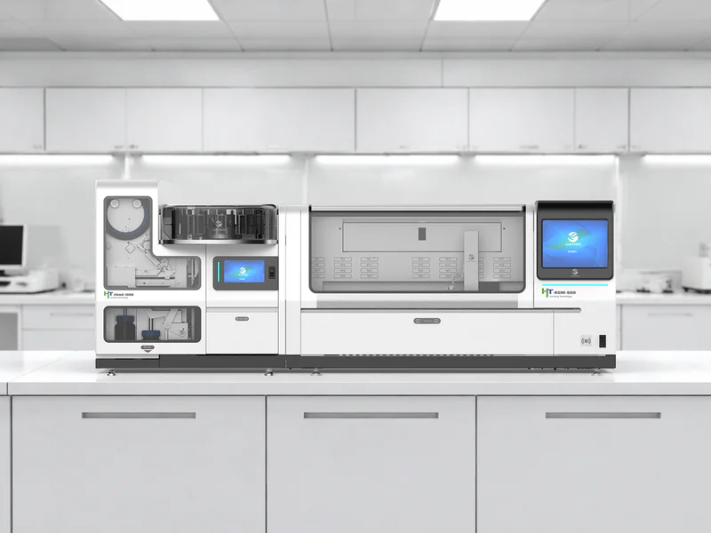
Every failure above maps to a design decision on our integrated staining and coverslipping workstation — the HT-BSHE-660 stainer and HT-FilmC-1000 coverslipper running as one line, from stain bath to finished slide with no human carry between.
The open-air window. The line transfers each finished slide straight into the coverslipper the moment staining ends. There is no rack to carry, so there is no wait to manage.
The beat. The coverslipper runs at 1,000 slides an hour against the stainer's 660 — deliberate headroom, so the last step never becomes the bottleneck. Finished slides land in a 240-slide output magazine, which is what lets the line finish a day's routine work and run unattended. Film is what lets the coverslipper keep that pace; whether film's per-slide cost fits a given lab is a separate question — price the tape before you buy a coverslipper.
Mixed workflows. Twelve protocols run simultaneously across six configurable staining stations, so H&E, Pap and frozen sections stay on the machine together instead of waiting in line. Reagent baths come in 750 ml and 260 ml sizes to match heavy and light stations.
First-to-last consistency. Reagent bottles carry RFID tags, so the machine knows each reagent's age and use. A patented adaptive protocol (ASBT) recalculates staining times by slide count and reagent condition while the run is in progress — the machine corrects for aging chemistry instead of letting it show up in the fourth rack. The H&E line uses five matched reagents — Mordant, Hematoxylin, Eosin, Differentiator and Bluing Reagent — tuned so the last slide of a maximum run stains like the first.
The room around it. The stainer sits within 125×73×65 cm and the coverslipper within 72×60×75 cm, and the whole line draws no more than 2,000 W — sized for an existing histology room, not a new one. The company runs ISO 9001 and ISO 13485, and the products carry CE. JunTeng integrated staining and coverslipping system
Questions Distributors Ask Me
"How do I sell this?" Lead with the four o'clock window, not with slides per hour. The buyer is the lab manager who knows exactly where their re-stains come from; the pitch is the gap that closes, and the rack consistency they can defend in an audit. For a lab doing two racks a day, a separate stainer and a technician with a coverslip bottle is still the right answer — say so, and the trust carries into the labs that have outgrown it.
"What do I write into a tender?" Put the architecture into the technical requirements: integrated automatic transfer from staining to coverslipping, coverslipping throughput above staining throughput, reagent-level monitoring with adaptive protocol adjustment, and unattended batch finishing. Those four lines describe a quality system, not a pair of machines.